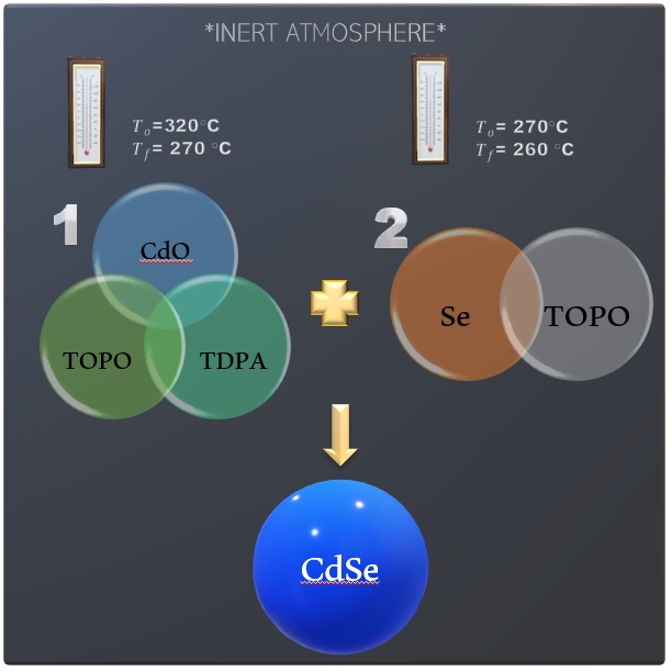
Figure 1. Diagram of Schlenk sintesis for QDs core CdSe [6].
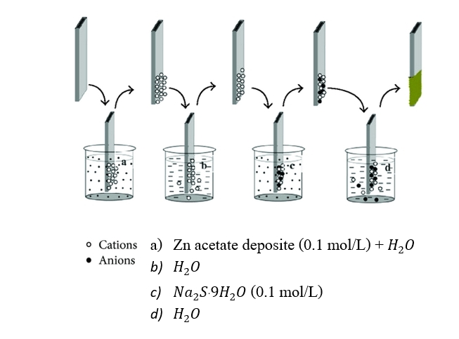
Figure 2. Diagram of SILAR technique for QDs shell Zn [7].
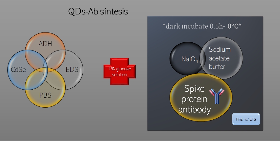
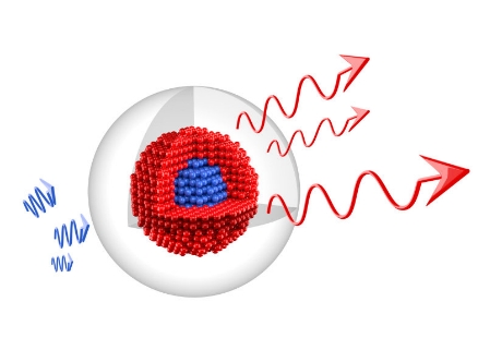
Figure 3. Diagram of conjugation of spike protein antibodies to QDs via oxidized Fc-carbohydrate groups [8].
Nanotechnology can be exploited to improve the utility of fluorescent markers used for diagnostic purposes.
They can be filled with a large number of imaging particles such as optically active compounds and radionuclides for the detection
with imaging equipment. Although fluorescent markers are routinely used in basic research and clinical diagnostic applications,
there are several inherent disadvantages with current techniques, including the requirement of color-matched lasers, the fluorescence bleaching,
and the lack of discriminatory capacity of multiple dyes.
Fluorescent nanocrystals potentially overcome these issues [2].
.
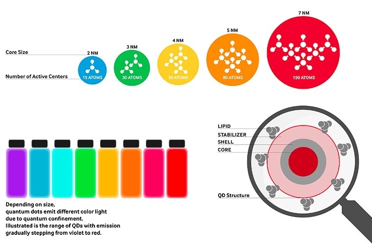
• QDs are used in LEDs and solid state lightning, displays and photovoltaics owing to their bright colors and their ability to emit rainbow of colors coupled with their high efficiencies, longer lifetimes and high extinction coefficient.
1) Sanjeeb, K. and Vinod, L. (2003) Nanotech approaches to drug delivery and imaging. Elsevier Science. 24, 1116-1117
2
Example
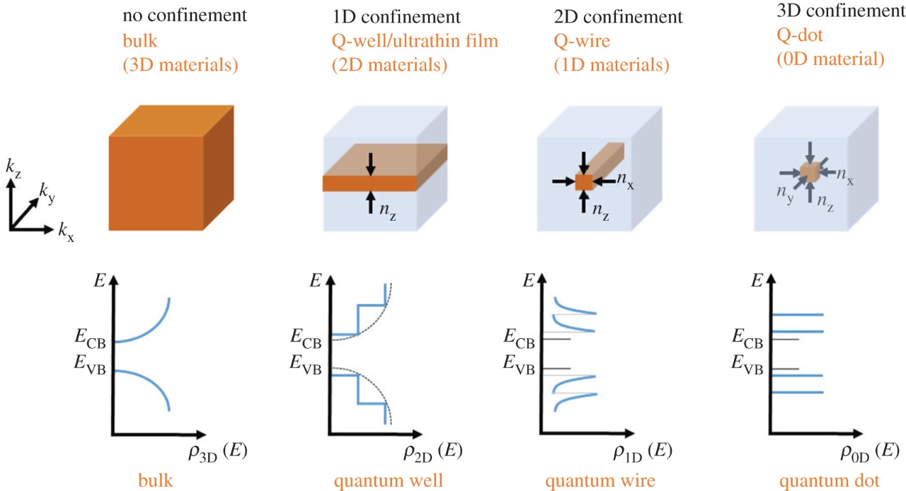
• Another application for QDs is owing to being zero dimensional and having a sharper density of states than higher dimensional structures and it is including transistors, solar cells, ultrafast all-optical switches and logic gates, among others.
• The particular size of QDs allow them to go anywhere in the body what makes them suitable for different biomedical applications like imaging and biosensors.
References
2) Chan, W.C. et al. (2002) Luminescent quantum dots for multiplexed biological detection and imaging. Curr. Opin. Biotechnol. 13, 40–46
3) Mitchell, P. (2001) Turning the spotlight on cellular imaging. Nat. Biotechnol. 19, 1013–1017
4) Mohs, A. and Provenzale, J. (2010) Applications of nanotechnology to imaging and therapy of brain tumors. Neuroimaging Clin N Am 20(3), 283–292
5) Shih, W., Shih, W., Li, H., Schillo, M. (2009) Water soluble quantum dots. Google Patents.
6) Luengo, J. et. al. (2017) Estudio de mojabilidad y caracterización del sistema pdms - vidrio en dispositivos microfluídicos. AFA 28, 10-14.
7)Asim N., et.al. (2014) Research and development aspects on chemical preparation techniques of photoanodes for dye sensitized solar cells. International Journal of Photoenergy.
8)Feng, W., et. al. (2016) Ultra Sensitive Detection of Influenza A Virus Based on Cdse/Zns Quantum Dots Immunoassay. SOJ Biochemistry.